
STEMPack Spectrum Imaging
A powerful method of obtaining detailed analytic data from a sample on an electron microscope equipped with scanning mode.

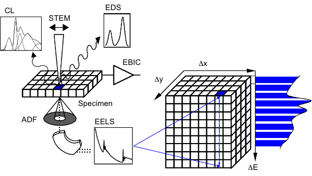
The STEMPack™ system enables a powerful method of obtaining detailed analytic data from a sample on an electron microscope equipped with scanning mode. By acquiring multimodal data in scanning mode, a wealth of information from precise positions within a scanned image or line profile is acquired. This approach is the basis of a family of techniques known as spectrum imaging (SI).
Spectrum imaging systematically probes a defined specimen area to gather the maximum possible information in a fully automated process. The STEMPack system supports the acquisition of data from a broad range of detectors, including: electron energy loss (EELS), characteristic x-ray emission (EDS), cathodoluminescence emission (CL), absorbed / induced current (EBAC/EBIC), and electron diffraction. The resulting spectral data is then analyzed with powerful processing and visualization methods to reveal unique detail in the specimen. The combination of detailed spectral information with high spatial resolution is the most powerful aspect of electron microscopy and the STEMPack system can help you realize this power.
- Maximize data flexibility and artifact rejection with feature rich acquisition options
- Use a single application to analyze dense data sets as simply as a single spectrum; while directly cross reference data and results
- Acquire the data size and region you choose with fully configurable acquisition
- Jointly acquire virtually any signal produced in the electron microscope as part of a spectrum image
- Obtain high speed and dose-efficient data acquisition for detail rich mapping using direct hardware communication
- Both new users and seasoned practitioners can achieve outstanding results with simple, yet flexible user interface

